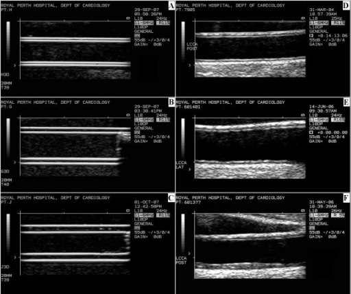
A TIPIC Ultrasonographic B-Mode Imaging of the Common Carotid
$ 9.99 · 4.9 (548) · In stock
Introduction/Patient Description Extracranial carotid duplex ultrasonography (DUS) was requested within 2 weeks after sudden onset of unilateral, evolving, neck pain. Signs and symptoms related to a 53 year-old man included local swelling, skin changes, increased, local sensations, and high sensitivity to palpation. Atherosclerotic risk factors were not noted. He had contralateral radiation therapy, neck and
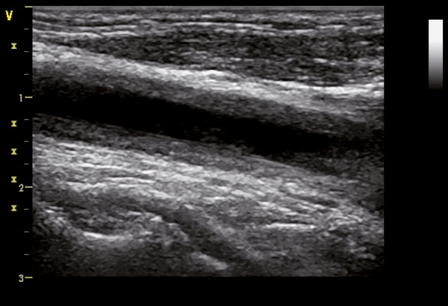
Figure 031_4969. B-mode ultrasonography of the common carotid artery in a 32-year-old woman
Beat-to-Beat Blood Pressure and Two-dimensional (axial and radial) Motion of the Carotid Artery Wall: Physiological Evaluation of Arterial Stiffness

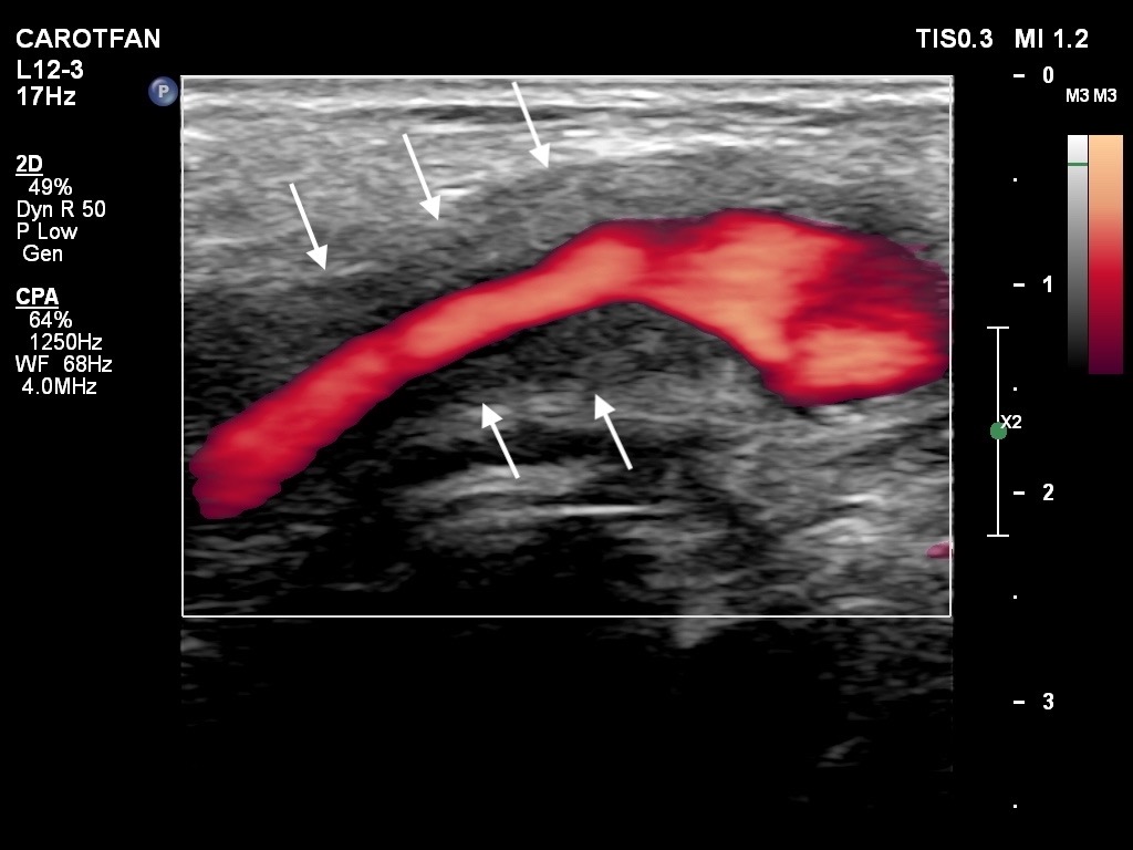
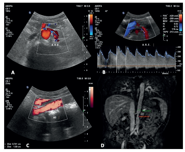
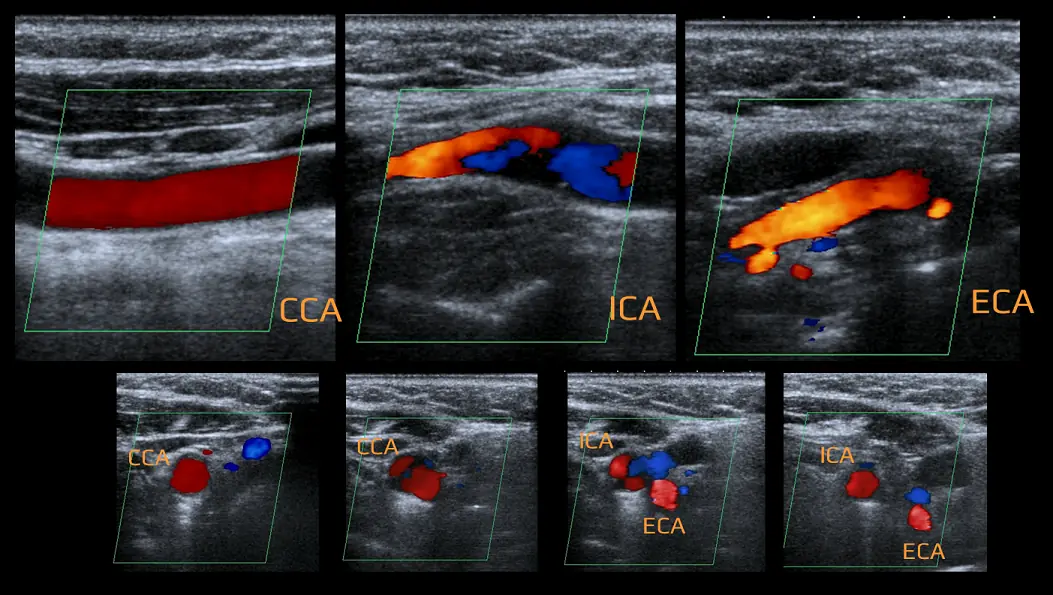
Internal carotid artery chronic occlusion: B-mode and colour Doppler flow appearance.Carotid Doppler
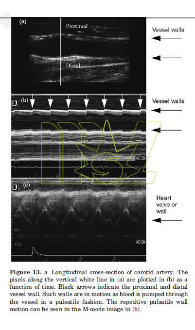
M Mode Ultrasound Definition
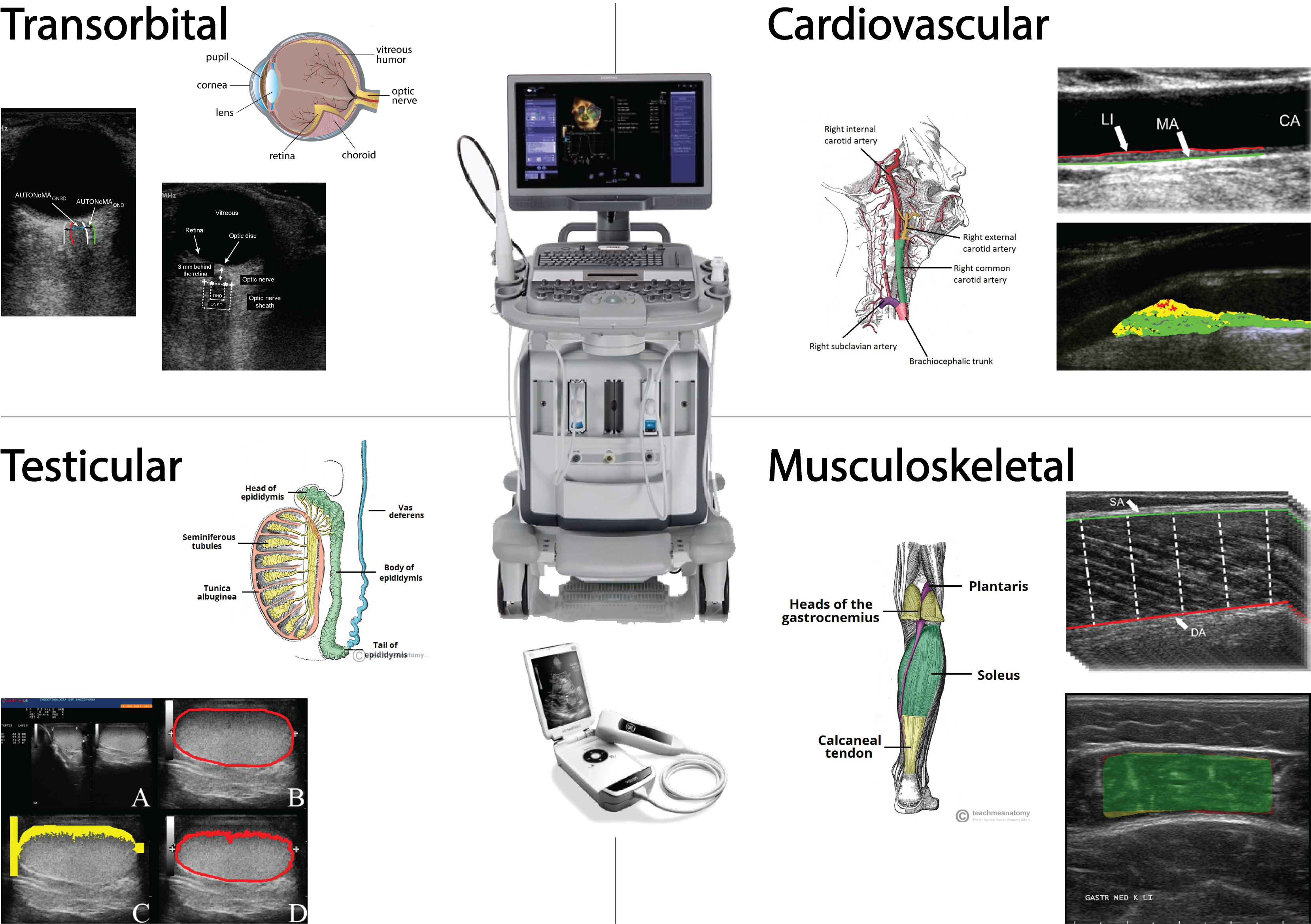
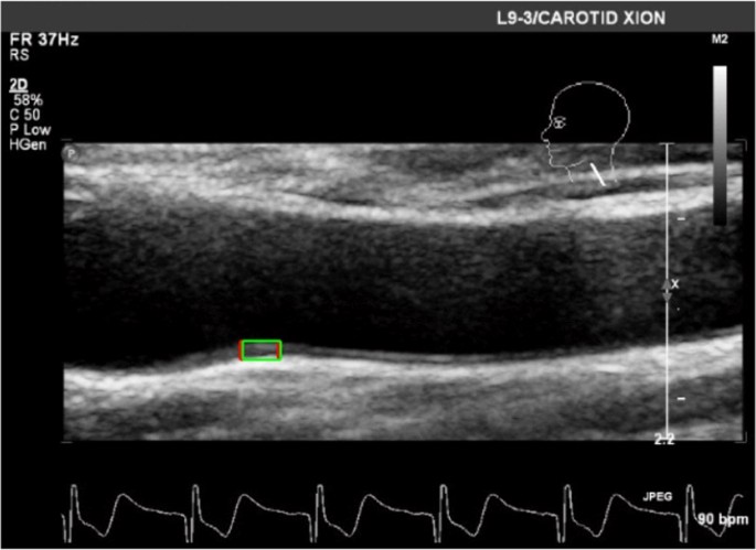
Ultrasound B-mode image segmentation and radiomics – BIOLAB@PoliTO
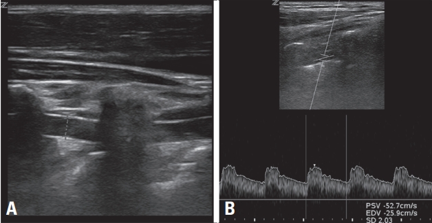
Carotid duplex ultrasound: interpretations and clinical applications
A TIPIC Ultrasonographic B-Mode Imaging of the Common Carotid

B-mode ultrasound image of the common carotid artery (longitudinal

Grey scale imaging (ultrasound), Radiology Reference Article
B-mode ultrasound images of phantom arteries. A. Phanto
![]()
4. B mode imaging – Carotid artery
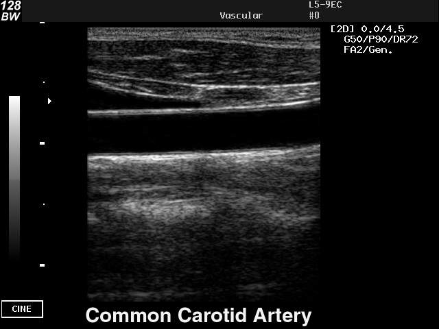
Ultrasound images • Common carotid artery, B-mode, echogramm №41
Extracranial Doppler Sonography