Figure 3 from Descriptive anatomy of the interscalene triangle and
$ 28.50 · 4.7 (178) · In stock

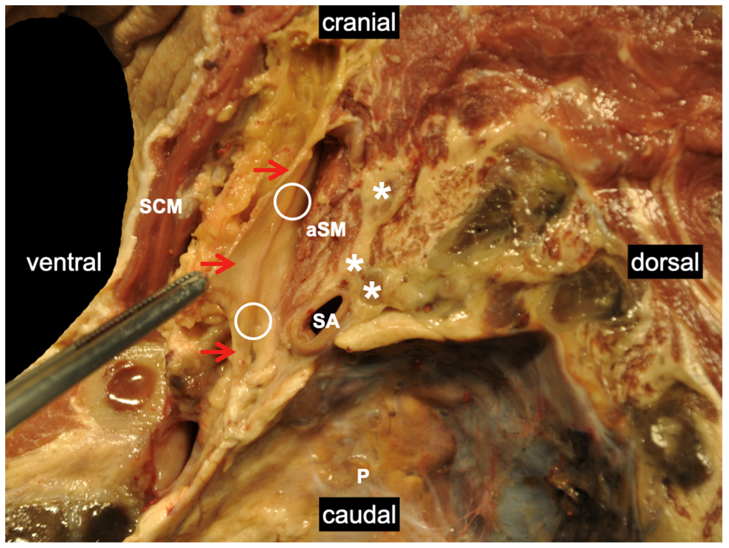
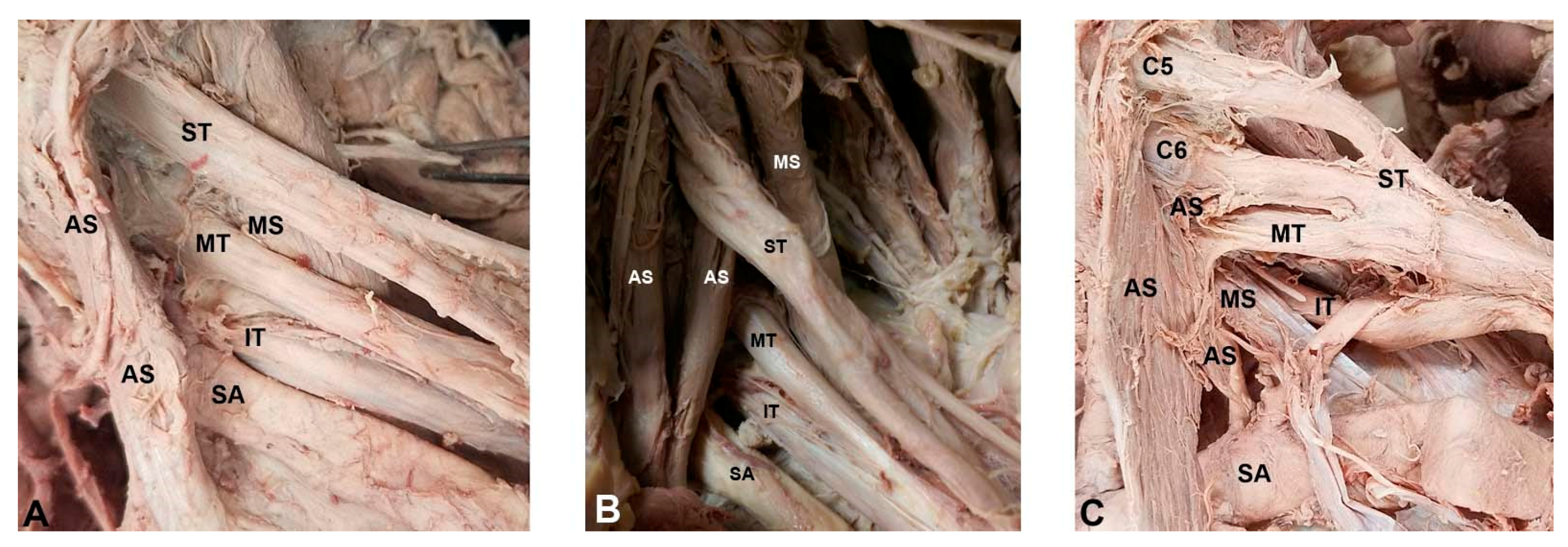
Fig 3. Depiction of the costoclavicular space. The neurovascular elements of the costoclavicular space can be seen here traveling superior to the first rib and inferior to the clavicle. The arrow indicates where measurements were taken. - "Descriptive anatomy of the interscalene triangle and the costoclavicular space and their relationship to thoracic outlet syndrome: a study of 60 cadavers."
Medicina, Free Full-Text
Robotic Surgery for Thoracic Outlet Syndrome
Thoracic Outlet Syndrome

Assessment of the interscalene triangle with different imaging

a: topography of the SSN in the suprascapular region. Area 1
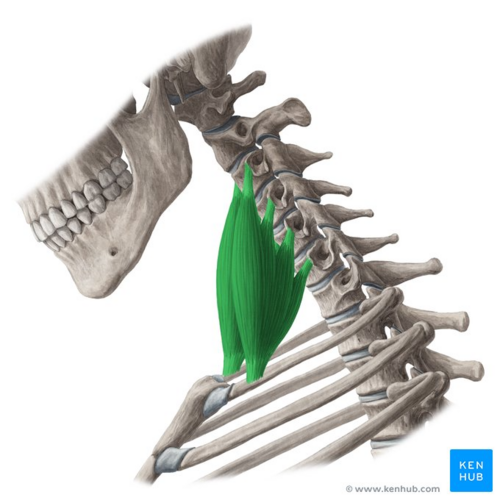
Scalene - Physiopedia

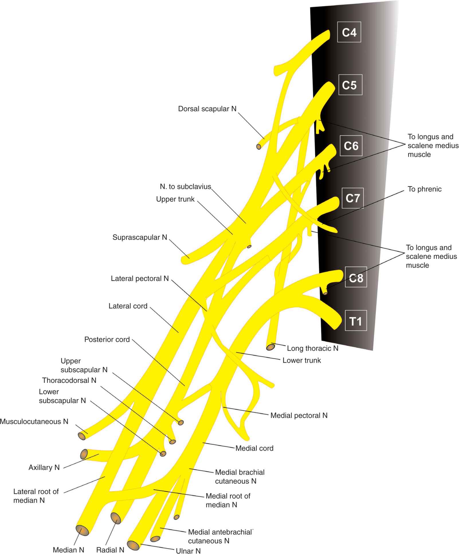
3 - Diagram: interscalene triangle and related structures Diagram
/images/vimeo_thumbnails/297907323/5rW9QulJRmGVbpv5dLLgjw_overlay.jpg)
Triangles of the neck: Anatomy, borders and contents
Diagnostics, Free Full-Text

Figure 3 from Descriptive anatomy of the interscalene triangle and the costoclavicular space and their relationship to thoracic outlet syndrome: a study of 60 cadavers.
Interscalene Brachial Plexus Block

Ultrasound guided interscalene block: Pro/Con